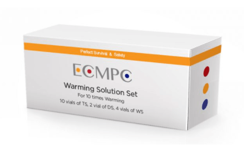
ECMPC - Vitrification Media
We develop the solutions that are necessary in the vitrification of mammalian embryos of any species, as well as in cryopreservation of oocytes and sperm of mammals.
In ECMPC we work with the most effective worldwide formula, counting with endorsed certificates with more than 15 years of experience supporting our services, That is why we are committed to each of our clients in providing not only effectiveness, but quality and commitment.
SHIPPING: Overnight Shipping Required to keep media at temperature - 2-8 degrees celsius.
MATERIALS
Vitrification Method Warming Kit
– Warming Solution (TS) :1 vial of 1.8mℓ
– Diluent Solution (DS) :1 vial of 0.5mℓ
– Washing Solution (WS) :1 vial of 1.0mℓ
– 1 Warming Plate with 4 wells
- Microscope (Turn off the heating plate)
- Stop watch (With count up function)
- Tweezers
- Micro pipette for 300μℓ
PREPARATION
Keep ES and VS vials at room temperature (25℃ to 27℃) for at least 2 hours before vitrification.
WARMING (1 Minute)
See Image Below
1.- Take the TS vial out of the incubator, and expel all of the solution out of it into the TS well (1.8ml, Fig. 8, Step1/①)
2.- Quickly (within 1 sec) put the Vitrification method from liquid nitrogen into the TS well (Fig. 8, Step1/②). Start counting up by the stop watch for 1 min.
3.- The oocyte/embryo releases from the Vitrification method sheet by itself, and begins to float.

VITRIFICATION1 (30 - 40 SECONDS)
1.- Aspirate the oocyte/embryo first, followed by 3mm of the TS into the pipette (Fig. 9, 1).
2.- Introduce the TS to the bottom of the DS well (Fig. 9, 2), then expel the oocyte/embryo slowly to the bottom of TS layer in DS well (Fig. 9, 3), and wait for 3 min (Fig. 8, Step 2/①).
3.- While waiting, fill the WS1 and the WS2 well with 300μℓ each of ws Solution (Fig. 8, Step 3/①).


STEP 1 STEP 2 STEP 3
WASHING (5 MINUTES)
1.- Aspirate the oocyte/embryo followed by 3mm of the DS into the pipette (Fig. 10,1).
2.- Introduce the DS to the bottom of the WS1 (Fig. 10, 2), and expel the oocyte/embryo slowly to the bottom of the DS layer in WS1 well (Fig. 10.3). Observe the shape of the oocyte/embryo and memorize it. Turn off the light, and wait for more than 3 min.
3.- After 3 min, compare the shape of the oocyte/embryo to the one memorized. Give a survival judgment if the shrinkage of the oocyte is recovered.
4.- Wait for 5 min in total (Fig. 8, Step3/②).
WASHING 2 (1 MIN)
1.- Aspirate the oocyte/embryo with minimal volume of the WS1.
2.- Put the oocyte/embryo on the surface of the WS2 well (Fig. 8, Step3/③).
3.- After the oocyte/embryo sinks to the bottom, aspirate and place it on the surface of a different location with in W2. Put the oocyte/embryo into the droplet of the culture media until ICSI or ET is performed.
4.- (Two to four hours culture for ICSI, and 3 hours for blastocyst transfer are recommended)
This media is not yet 510K Cleared.


